Tuesday Poster Session
Category: GI Bleeding
P3515 - A Case of Meckels Diverticulum in a 25-Year-Old Patient With Recurrent Hematochezia
Tuesday, October 24, 2023
10:30 AM - 4:00 PM PT
Location: Exhibit Hall

Has Audio
- LG
Lasha Gogokhia, MD
New York-Presbyterian Hospital/Weill Cornell Medical Center
New York, New York
Presenting Author(s)
Johannes Untch, 1, Lasha Gogokhia, MD2, David Wan, BS, MD3
1Technische Universität München, School of Medicine, München, Bayern, Germany; 2New York-Presbyterian Hospital/Weill Cornell Medical Center, New York, NY; 3Weill Cornell Medicine, New York, NY
Introduction: Meckel's diverticulum (MD) is the most common hereditary malformation in the gastrointestinal tract presenting in 2% of the population. It stems from a failure of the Vitelline duct to obliterate during embryogenesis, contains all coats of the small bowel wall and the mucosa can contain ectopic gastric or pancreatic tissue. MD rarely become clinically symptomatic, but complications include hemorrhage, intussusception, intestinal stricture, obstruction, and perforation.
Case Description/Methods: A 25-year-old previously healthy woman was referred to the emergency room (ED) for symptomatic iron deficiency anemia (Hb 6.2 g/dL). Rectal exam demonstrated melenic stools. Next day she had episodes of hematochezia requiring multiple unit blood transfusions to stabilize her Hb. Complete luminal evaluation with esophagogastroduodenoscopy, colonoscopy and video capsule endoscopy (VCE) demonstrated fresh blood in colon but was without identification of active bleeding source along the examined GI tract. Nuclear medicine gastrointestinal bleeding scan using Tc99m-labelled RBCs was negative at that time as well and the patient was discharged. Seven months later the patient had three hospital admissions for hematochezia with acute anemia during which her complete endoscopic evaluation did not reveal an active bleeding source. Repeat CT abdomen pelvis showed mucosal hyperenhancement and slight surrounding inflammation but no active hemorrhage suggestive of MD which was later confirmed with Tc99m-SPECT/CT (Meckel Scan). The patient underwent laparoscopic small bowel resection with unremarkable post-op course and has since had no more episodes of bleeding.
Discussion: MD can be difficult to diagnose because of its rarity, non-specific imaging findings and clinical presentation that resembles a variety of other GI conditions. In this case, presentation with hematochezia led to the initial workup including EGD/colonoscopy, VCE and nuclear medicine bleeding scan that could not identify an active bleeding source. Only repeat CT and Meckel scan led to the final diagnosis after recurrent bleeding episodes. Meckel scan is the most accurate non-invasive test for MD and its sensitivity in children can be over 90%, however in adults this number is significantly lower and repeat scans may be necessary. Although rare, MD should be considered on differential in young patients with obscure gastrointestinal bleeding and specific imaging modality should be obtained to rule out congenital malformations.

Disclosures:
Johannes Untch, 1, Lasha Gogokhia, MD2, David Wan, BS, MD3. P3515 - A Case of Meckels Diverticulum in a 25-Year-Old Patient With Recurrent Hematochezia, ACG 2023 Annual Scientific Meeting Abstracts. Vancouver, BC, Canada: American College of Gastroenterology.
1Technische Universität München, School of Medicine, München, Bayern, Germany; 2New York-Presbyterian Hospital/Weill Cornell Medical Center, New York, NY; 3Weill Cornell Medicine, New York, NY
Introduction: Meckel's diverticulum (MD) is the most common hereditary malformation in the gastrointestinal tract presenting in 2% of the population. It stems from a failure of the Vitelline duct to obliterate during embryogenesis, contains all coats of the small bowel wall and the mucosa can contain ectopic gastric or pancreatic tissue. MD rarely become clinically symptomatic, but complications include hemorrhage, intussusception, intestinal stricture, obstruction, and perforation.
Case Description/Methods: A 25-year-old previously healthy woman was referred to the emergency room (ED) for symptomatic iron deficiency anemia (Hb 6.2 g/dL). Rectal exam demonstrated melenic stools. Next day she had episodes of hematochezia requiring multiple unit blood transfusions to stabilize her Hb. Complete luminal evaluation with esophagogastroduodenoscopy, colonoscopy and video capsule endoscopy (VCE) demonstrated fresh blood in colon but was without identification of active bleeding source along the examined GI tract. Nuclear medicine gastrointestinal bleeding scan using Tc99m-labelled RBCs was negative at that time as well and the patient was discharged. Seven months later the patient had three hospital admissions for hematochezia with acute anemia during which her complete endoscopic evaluation did not reveal an active bleeding source. Repeat CT abdomen pelvis showed mucosal hyperenhancement and slight surrounding inflammation but no active hemorrhage suggestive of MD which was later confirmed with Tc99m-SPECT/CT (Meckel Scan). The patient underwent laparoscopic small bowel resection with unremarkable post-op course and has since had no more episodes of bleeding.
Discussion: MD can be difficult to diagnose because of its rarity, non-specific imaging findings and clinical presentation that resembles a variety of other GI conditions. In this case, presentation with hematochezia led to the initial workup including EGD/colonoscopy, VCE and nuclear medicine bleeding scan that could not identify an active bleeding source. Only repeat CT and Meckel scan led to the final diagnosis after recurrent bleeding episodes. Meckel scan is the most accurate non-invasive test for MD and its sensitivity in children can be over 90%, however in adults this number is significantly lower and repeat scans may be necessary. Although rare, MD should be considered on differential in young patients with obscure gastrointestinal bleeding and specific imaging modality should be obtained to rule out congenital malformations.

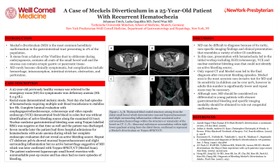
Figure: Figure 1. A./B. Thickened blind-ended structure arising from the distal small bowel which demonstrates mucosal hyperenhancement and slight surrounding inflammation without associated active extravasation/hemorrhage within the structure or remainder of the bowel on CT.
C./D.: Abnormal radiotracer accumulation within right lower quadrant arising from the distal ileum, confirmed to represent a Meckel's diverticulum on Tc99m-SPECT/CT.
C./D.: Abnormal radiotracer accumulation within right lower quadrant arising from the distal ileum, confirmed to represent a Meckel's diverticulum on Tc99m-SPECT/CT.
Disclosures:
Johannes Untch indicated no relevant financial relationships.
Lasha Gogokhia indicated no relevant financial relationships.
David Wan indicated no relevant financial relationships.
Johannes Untch, 1, Lasha Gogokhia, MD2, David Wan, BS, MD3. P3515 - A Case of Meckels Diverticulum in a 25-Year-Old Patient With Recurrent Hematochezia, ACG 2023 Annual Scientific Meeting Abstracts. Vancouver, BC, Canada: American College of Gastroenterology.
