Tuesday Poster Session
Category: GI Bleeding
P3516 - A Bleeding Duodenal Lipoma: An Uncommon Diagnosis
Tuesday, October 24, 2023
10:30 AM - 4:00 PM PT
Location: Exhibit Hall

Has Audio

Aastha V. Bharwad, MD
University of Kansas School of Medicine
Wichita, KS
Presenting Author(s)
Aastha Bharwad, MD1, Lawrence Zhou, MD1, Wael TAWFIC. Mohamed, MD2, William J.. Salyers, MD, MPH1, Thao N. Nguyen, DO3
1University of Kansas School of Medicine, Wichita, KS; 2University of Missouri-Kansas City, Kansas City, MO; 3Ascension Medical Group, Wichita, KS
Introduction: Gastrointestinal (GI) lipomas are relatively uncommon benign tumors and when present, occur commonly in the colon. Bleeding duodenal lipomas (DL) are very rare and challenging to manage. We present a case of large pedunculated duodenal lipoma, that was submucosally resected successfully, achieving adequate hemostasis.
Case Description/Methods: A 44-year-old male with a past medical history of hyperlipidemia, attention deficit hyperactivity disorder, obstructive sleep apnea, and depression presented to the emergency room with two days of black stools. He denied alcohol or non-steroidal anti-inflammatory drug use. His hemoglobin (Hgb) on admission was 7.8 g/dL, and due to active bleeding, he received two units of blood and one dose of iron dextran. During admission, esophagogastroduodenoscopy (EGD) showed no active bleeding, non-obstructing Schatzki's ring measuring 3 cm, a small hiatal hernia, and non-bleeding erosive gastritis in the antrum. Colonoscopy did not reveal any abnormalities. He was eventually discharged on proton pump inhibitor and ferrous sulfate with a Hgb of 8.3 g/dL and follow up with outpatient pill endoscopy.
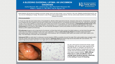
Pill endoscopy showed no old or fresh blood in the stomach, red fresh blood with active oozing in the distal duodenum, and dark blood throughout the small bowel, but inability to evaluate the colon due to poor preparation. Follow-up push enteroscopy (Figure 1) revealed a large broad-based pedunculated polyp with a broad stalk in the fourth portion of duodenum with a nonbleeding clean-based ulcer present at the inferior aspect of the polyp. Initial biopsy of the lesion was unremarkable.
Subsequent EGD two weeks later for endoscopic mucosal resection (EMR) successfully removed the single 25 mm pedunculated polyp via ligation with a PolyLoop at base of the lesion followed by hot snare resection. Pathology (Figure 2) showed benign lipoma associated with focal ulceration. The patient’s Hgb post-procedure improved to 12.2 mg/dL with no further episodes of dark stools.
Discussion: DL are a rare source of acute and chronic upper GI bleeding, with very few cases reported so far. Mucosal erosion or ulceration over lipoma can lead to severe bleeding. Symptomatic tumors are usually removed endoscopically, and larger ones may require surgical resection. EMR utilizing PolyLoop is a safe and effective way to manage large duodenal lipomas, especially in the setting of recent GI bleeding.

Disclosures:
Aastha Bharwad, MD1, Lawrence Zhou, MD1, Wael TAWFIC. Mohamed, MD2, William J.. Salyers, MD, MPH1, Thao N. Nguyen, DO3. P3516 - A Bleeding Duodenal Lipoma: An Uncommon Diagnosis, ACG 2023 Annual Scientific Meeting Abstracts. Vancouver, BC, Canada: American College of Gastroenterology.
1University of Kansas School of Medicine, Wichita, KS; 2University of Missouri-Kansas City, Kansas City, MO; 3Ascension Medical Group, Wichita, KS
Introduction: Gastrointestinal (GI) lipomas are relatively uncommon benign tumors and when present, occur commonly in the colon. Bleeding duodenal lipomas (DL) are very rare and challenging to manage. We present a case of large pedunculated duodenal lipoma, that was submucosally resected successfully, achieving adequate hemostasis.
Case Description/Methods: A 44-year-old male with a past medical history of hyperlipidemia, attention deficit hyperactivity disorder, obstructive sleep apnea, and depression presented to the emergency room with two days of black stools. He denied alcohol or non-steroidal anti-inflammatory drug use. His hemoglobin (Hgb) on admission was 7.8 g/dL, and due to active bleeding, he received two units of blood and one dose of iron dextran. During admission, esophagogastroduodenoscopy (EGD) showed no active bleeding, non-obstructing Schatzki's ring measuring 3 cm, a small hiatal hernia, and non-bleeding erosive gastritis in the antrum. Colonoscopy did not reveal any abnormalities. He was eventually discharged on proton pump inhibitor and ferrous sulfate with a Hgb of 8.3 g/dL and follow up with outpatient pill endoscopy.
Pill endoscopy showed no old or fresh blood in the stomach, red fresh blood with active oozing in the distal duodenum, and dark blood throughout the small bowel, but inability to evaluate the colon due to poor preparation. Follow-up push enteroscopy (Figure 1) revealed a large broad-based pedunculated polyp with a broad stalk in the fourth portion of duodenum with a nonbleeding clean-based ulcer present at the inferior aspect of the polyp. Initial biopsy of the lesion was unremarkable.
Subsequent EGD two weeks later for endoscopic mucosal resection (EMR) successfully removed the single 25 mm pedunculated polyp via ligation with a PolyLoop at base of the lesion followed by hot snare resection. Pathology (Figure 2) showed benign lipoma associated with focal ulceration. The patient’s Hgb post-procedure improved to 12.2 mg/dL with no further episodes of dark stools.
Discussion: DL are a rare source of acute and chronic upper GI bleeding, with very few cases reported so far. Mucosal erosion or ulceration over lipoma can lead to severe bleeding. Symptomatic tumors are usually removed endoscopically, and larger ones may require surgical resection. EMR utilizing PolyLoop is a safe and effective way to manage large duodenal lipomas, especially in the setting of recent GI bleeding.

Figure: Figure 1: Push enteroscopy showing a single 25 mm pedunculated polyp with a broad stalk in the fourth portion of the duodenum
Figure 2: Pathology revealing benign lipoma
Figure 2: Pathology revealing benign lipoma
Disclosures:
Aastha Bharwad indicated no relevant financial relationships.
Lawrence Zhou indicated no relevant financial relationships.
Wael Mohamed indicated no relevant financial relationships.
William Salyers indicated no relevant financial relationships.
Thao Nguyen indicated no relevant financial relationships.
Aastha Bharwad, MD1, Lawrence Zhou, MD1, Wael TAWFIC. Mohamed, MD2, William J.. Salyers, MD, MPH1, Thao N. Nguyen, DO3. P3516 - A Bleeding Duodenal Lipoma: An Uncommon Diagnosis, ACG 2023 Annual Scientific Meeting Abstracts. Vancouver, BC, Canada: American College of Gastroenterology.
