Tuesday Poster Session
Category: Esophagus
P3285 - An Unusual Diagnosis of a Tracheoesophageal Fistula After Esophageal Stent Placement
Tuesday, October 24, 2023
10:30 AM - 4:00 PM PT
Location: Exhibit Hall

Has Audio

Seth Lipshutz, DO
Jefferson Health Northeast
Philadelphia, PA
Presenting Author(s)
Seth Lipshutz, DO1, Babak Etemad, MD2, Bryan Stone, DO3, Julian Remouns, DO2, Victoria Burris, DO4
1Jefferson Health Northeast, Philadelphia, PA; 2Lankenau Medical Center, Philadelphia, PA; 3Lankenau Medical Center, Wynnewood, PA; 4MRHC, Philadelphia, PA
Introduction: Esophageal cancer is often diagnosed at an advanced stage, with many patients found to have regional or metastatic disease at time of diagnosis. Patients often present with frequent coughing after oral intake, recurrent episodes of aspiration, or unexplained malnutrition, often requiring palliative therapies, such as esophageal stents. These stents are important tools for palliative treatment of inoperable esophageal malignancies and can be complicated by broncho-esophageal fistulas (BEFs) and tracheoesophageal fistulas (TEFs) connecting the trachea or bronchi to the esophagus.1 Although these fistulas generally occur due to damage from primary malignancy, fistulas after stent placement can uncommonly occur, and prompt identification and treatment is imperative.
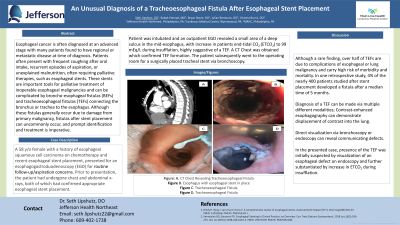
Case Description/Methods: A 58 y/o female with PMHx of esophageal squamous cell carcinoma on chemotherapy, with recent esophageal stent placement, presented for esophagogastroduodenoscopy (EGD), and was admitted for concern for tracheoesophageal fistula. Prior to presentation, the patient had undergone chest and abdominal x-rays, both of which had confirmed appropriate esophageal stent placement. During admission, the patient underwent EGD, where there was concern for TEF formation. Per operative note, there was a small area with suggestion of a deeper sulcus, with increase in patients CO2 to 99 during insufflation, highly suggestive of a TEF. Following EGD, patient underwent CT Chest, which revealed TEF formation. The patient subsequently went to the operating room for a surgically placed tracheal stent via bronchoscopy.
Discussion: Although a rare finding, over half of TEFs are due to complications of esophageal or lung malignancy and carry high risk of morbidity and mortality. In one retrospective study, 4% of the nearly 400 patients studied after stent placement developed a fistula after a median time of 5 months.1 Diagnosis of a TEF can be aided by obtaining contrast-enhanced esophagography that demonstrates displacement of contrast into the lung, or by direct visualization via bronchoscopy or endoscopy. The case we present was found to have a diagnosis of fistula by way of multiple modalities, in addition to clinical evaluation during EGD with increase in CO2 to 99, concerning for fistula formation into the trachea.
References:
1. Patel H, et al, The Unexpected Formation of a Broncho-Esophageal Fistula of the Right Main Stem Bronchus Status Post Esophageal Stent Placement: A Case Report. Cureus. 2022 Jan 26;14(1)

Disclosures:
Seth Lipshutz, DO1, Babak Etemad, MD2, Bryan Stone, DO3, Julian Remouns, DO2, Victoria Burris, DO4. P3285 - An Unusual Diagnosis of a Tracheoesophageal Fistula After Esophageal Stent Placement, ACG 2023 Annual Scientific Meeting Abstracts. Vancouver, BC, Canada: American College of Gastroenterology.
1Jefferson Health Northeast, Philadelphia, PA; 2Lankenau Medical Center, Philadelphia, PA; 3Lankenau Medical Center, Wynnewood, PA; 4MRHC, Philadelphia, PA
Introduction: Esophageal cancer is often diagnosed at an advanced stage, with many patients found to have regional or metastatic disease at time of diagnosis. Patients often present with frequent coughing after oral intake, recurrent episodes of aspiration, or unexplained malnutrition, often requiring palliative therapies, such as esophageal stents. These stents are important tools for palliative treatment of inoperable esophageal malignancies and can be complicated by broncho-esophageal fistulas (BEFs) and tracheoesophageal fistulas (TEFs) connecting the trachea or bronchi to the esophagus.1 Although these fistulas generally occur due to damage from primary malignancy, fistulas after stent placement can uncommonly occur, and prompt identification and treatment is imperative.
Case Description/Methods: A 58 y/o female with PMHx of esophageal squamous cell carcinoma on chemotherapy, with recent esophageal stent placement, presented for esophagogastroduodenoscopy (EGD), and was admitted for concern for tracheoesophageal fistula. Prior to presentation, the patient had undergone chest and abdominal x-rays, both of which had confirmed appropriate esophageal stent placement. During admission, the patient underwent EGD, where there was concern for TEF formation. Per operative note, there was a small area with suggestion of a deeper sulcus, with increase in patients CO2 to 99 during insufflation, highly suggestive of a TEF. Following EGD, patient underwent CT Chest, which revealed TEF formation. The patient subsequently went to the operating room for a surgically placed tracheal stent via bronchoscopy.
Discussion: Although a rare finding, over half of TEFs are due to complications of esophageal or lung malignancy and carry high risk of morbidity and mortality. In one retrospective study, 4% of the nearly 400 patients studied after stent placement developed a fistula after a median time of 5 months.1 Diagnosis of a TEF can be aided by obtaining contrast-enhanced esophagography that demonstrates displacement of contrast into the lung, or by direct visualization via bronchoscopy or endoscopy. The case we present was found to have a diagnosis of fistula by way of multiple modalities, in addition to clinical evaluation during EGD with increase in CO2 to 99, concerning for fistula formation into the trachea.
References:
1. Patel H, et al, The Unexpected Formation of a Broncho-Esophageal Fistula of the Right Main Stem Bronchus Status Post Esophageal Stent Placement: A Case Report. Cureus. 2022 Jan 26;14(1)

Figure: A. CT Chest Revealing Tracheoesophageal Fistula
B. Esophagus with esophageal stent in place
C. Tracheoesophageal Fistula
D. Tracheoesophageal Fistula
B. Esophagus with esophageal stent in place
C. Tracheoesophageal Fistula
D. Tracheoesophageal Fistula
Disclosures:
Seth Lipshutz indicated no relevant financial relationships.
Babak Etemad indicated no relevant financial relationships.
Bryan Stone indicated no relevant financial relationships.
Julian Remouns indicated no relevant financial relationships.
Victoria Burris indicated no relevant financial relationships.
Seth Lipshutz, DO1, Babak Etemad, MD2, Bryan Stone, DO3, Julian Remouns, DO2, Victoria Burris, DO4. P3285 - An Unusual Diagnosis of a Tracheoesophageal Fistula After Esophageal Stent Placement, ACG 2023 Annual Scientific Meeting Abstracts. Vancouver, BC, Canada: American College of Gastroenterology.
