Tuesday Poster Session
Category: Esophagus
P3291 - Not so Typical Appendicitis Leads to Diagnosis of Impressive Signet Ring Esophageal Adenocarcinoma
Tuesday, October 24, 2023
10:30 AM - 4:00 PM PT
Location: Exhibit Hall

Has Audio

Shane Quo, DO
University of Miami at Holy Cross Hospital
Fort Lauderdale, FL
Presenting Author(s)
Shane Quo, DO1, Mary Parianos, MD2, Eduardo Villa, MD1
1University of Miami Holy Cross Hospital, Fort Lauderdale, FL; 2University of MIami Holy Cross Hospital, Fort Lauderdale, FL
Introduction: The rates of esophageal cancer are increasing, and it is now the sixth leading cause of cancer deaths worldwide. Typically, esophageal cancer is either adenocarcinoma or squamous cell carcinoma. The majority of prior cases showed that squamous cell carcinoma was more prevalent than adenocarcinoma, however, those numbers have now drastically shifted. Adenocarcinoma arises from the mucus-secreting glands, while squamous develops from the flat cells that line the esophagus. The management of esophageal cancer depends on the Siewert classification, which is based on the location of the cancer. Our case highlights a unique presentation of esophageal cancer, diagnosed initially as appendicitis.
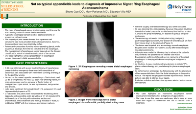
Case Description/Methods: A 62-year-old male with a history of hypertension presents with abdominal pain associated with vomiting and fatigue for the past 2-3 weeks. He reports decreased appetite and 20 lbs of weight loss. He has a smoking history, no prior colonoscopy, and no family history of cancer. Upon arrival, he was hypertensive, tachycardiac, and afebrile. CT abdomen and pelvis were remarkable for thickening of the appendix, 1.7cm mass in the liver, ascites, retroperitoneal lymphadenopathy. Due to imaging, antibiotics were started. Gastroenterology (GI) was consulted. GI was unable to perform a colonoscopy as the patient was not able to tolerate the bowel prep. XR esophagram ordered and results shown in Figure 1. GI decided on an upper endoscopy (EGD). The EGD showed an obstructing gastroesophageal junction tumor extending from the mid esophagus to gastric fundus. Pathology was notable for invasive, poorly differentiated signet ring adenocarcinoma.Repeat EGD was performed for esophageal stenting. The decision was made to start total parenteral nutrition. The plan initially included radiation therapy with the idea to attempt enteral nutrition in the future. However, due to the aggressive nature of this advanced malignancy, he would continue to decline and ultimately palliative care was decided.
Discussion: This case illustrates how crucial an appropriate medical history is essential for appropriate diagnosis of disease. The initial complaint was abdominal pain with vomiting in the setting of an inflamed appendix on imaging. However, the associated symptoms of significant weight loss, decreased appetite, and chills proved to be significant. We hope to use this case as a cautionary tale to keep an open mind regarding differentials and not to anchor onto a diagnosis.

Disclosures:
Shane Quo, DO1, Mary Parianos, MD2, Eduardo Villa, MD1. P3291 - Not so Typical Appendicitis Leads to Diagnosis of Impressive Signet Ring Esophageal Adenocarcinoma, ACG 2023 Annual Scientific Meeting Abstracts. Vancouver, BC, Canada: American College of Gastroenterology.
1University of Miami Holy Cross Hospital, Fort Lauderdale, FL; 2University of MIami Holy Cross Hospital, Fort Lauderdale, FL
Introduction: The rates of esophageal cancer are increasing, and it is now the sixth leading cause of cancer deaths worldwide. Typically, esophageal cancer is either adenocarcinoma or squamous cell carcinoma. The majority of prior cases showed that squamous cell carcinoma was more prevalent than adenocarcinoma, however, those numbers have now drastically shifted. Adenocarcinoma arises from the mucus-secreting glands, while squamous develops from the flat cells that line the esophagus. The management of esophageal cancer depends on the Siewert classification, which is based on the location of the cancer. Our case highlights a unique presentation of esophageal cancer, diagnosed initially as appendicitis.
Case Description/Methods: A 62-year-old male with a history of hypertension presents with abdominal pain associated with vomiting and fatigue for the past 2-3 weeks. He reports decreased appetite and 20 lbs of weight loss. He has a smoking history, no prior colonoscopy, and no family history of cancer. Upon arrival, he was hypertensive, tachycardiac, and afebrile. CT abdomen and pelvis were remarkable for thickening of the appendix, 1.7cm mass in the liver, ascites, retroperitoneal lymphadenopathy. Due to imaging, antibiotics were started. Gastroenterology (GI) was consulted. GI was unable to perform a colonoscopy as the patient was not able to tolerate the bowel prep. XR esophagram ordered and results shown in Figure 1. GI decided on an upper endoscopy (EGD). The EGD showed an obstructing gastroesophageal junction tumor extending from the mid esophagus to gastric fundus. Pathology was notable for invasive, poorly differentiated signet ring adenocarcinoma.Repeat EGD was performed for esophageal stenting. The decision was made to start total parenteral nutrition. The plan initially included radiation therapy with the idea to attempt enteral nutrition in the future. However, due to the aggressive nature of this advanced malignancy, he would continue to decline and ultimately palliative care was decided.
Discussion: This case illustrates how crucial an appropriate medical history is essential for appropriate diagnosis of disease. The initial complaint was abdominal pain with vomiting in the setting of an inflamed appendix on imaging. However, the associated symptoms of significant weight loss, decreased appetite, and chills proved to be significant. We hope to use this case as a cautionary tale to keep an open mind regarding differentials and not to anchor onto a diagnosis.

Figure: Figure 1. XR Esophagram performed due to decreased appetite and inability to tolerate bowel preparation
Disclosures:
Shane Quo indicated no relevant financial relationships.
Mary Parianos indicated no relevant financial relationships.
Eduardo Villa indicated no relevant financial relationships.
Shane Quo, DO1, Mary Parianos, MD2, Eduardo Villa, MD1. P3291 - Not so Typical Appendicitis Leads to Diagnosis of Impressive Signet Ring Esophageal Adenocarcinoma, ACG 2023 Annual Scientific Meeting Abstracts. Vancouver, BC, Canada: American College of Gastroenterology.
