Tuesday Poster Session
Category: Colon
P3064 - Rare Case of Colonic Amyloidosis
Tuesday, October 24, 2023
10:30 AM - 4:00 PM PT
Location: Exhibit Hall

Has Audio

Charles Yi, MD
University of South Alabama
Mobile, AL
Presenting Author(s)
Charles Yi, MD1, Karan B. Singh, MD1, Alvin Green, DO, MPH1, Xuebao Zhang, MD2, Travis Berry, DO3, Phillip Henderson, DO1
1University of South Alabama, Mobile, AL; 2University of South Alabama Health Systems, Mobile, AL; 3University of South Alabama College of Medicine, Mobile, AL
Introduction: Amyloidosis is a heterogeneous disorder with extracellular deposition of insoluble proteins. The most common presentation is primary light chain systemic amyloidosis with an incidence of 10 per million person-years. Less common is localized amyloidosis which accounts for only 15% of all amyloid cases. We present a case of localized gastrointestinal amyloidosis of the colon.
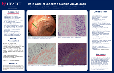
Case Description/Methods: This is a 67 year old female with history of mixed connective tissue disorder (Lupus with Sjogrens overlap) on immunosuppressive therapy, GERD, hypertrygliceridemia, CKD3 who presented to the GI clinic for chronic diarrhea and a positive AMA testing.
She described alternating episodes of diarrhea and constipation for a prolonged period of time, intermittent dysphagia to liquids, nausea, and self-resolving episodes of hematochezia without abdominal pain, emesis, odynophagia, or fevers. Previous endoscopy at another institution was noncontributory. EGD was performed revealing hemorrhagic gastropathy. Colonoscopy revealed polyps in the ascending colon and descending colon with inflammation and loss of vascular pattern in the sigmoid colon. Gastric biopsy results returned positive for H pylori for which the patient received quad therapy. Histopathology of the polyps and the sigmoid colon both showed diffuse vascular wall amyloid deposition highlighted by Congo red stain.
Subsequent workup with hematology/oncology revealed no AL amyloidosis involvement of the heart, kidneys, or bone marrow, and treatment was deferred as she remained stable while on observation receiving treatment for H pylori. Two years after the initial biopsy results, our patient returned to the clinic for worsening reflux symptoms now poorly controlled on her prior Nexium regimen. Repeat EGD and CSC performed showed normal biopsy of esophagus but signs of amyloidosis involving the duodenal bulb in addition to the entire colon.
Discussion: Amyloidosis represents a distinctively rare condition, and its localized manifestation in the gastrointestinal system is an even rarer occurrence. Despite their rarity, conditions like these are indeed extant and must be considered within the differential diagnoses by all clinicians. Management involves either observation in asymptomatic patients or ongoing surveillance for other gastrointestinal symptoms.

Disclosures:
Charles Yi, MD1, Karan B. Singh, MD1, Alvin Green, DO, MPH1, Xuebao Zhang, MD2, Travis Berry, DO3, Phillip Henderson, DO1. P3064 - Rare Case of Colonic Amyloidosis, ACG 2023 Annual Scientific Meeting Abstracts. Vancouver, BC, Canada: American College of Gastroenterology.
1University of South Alabama, Mobile, AL; 2University of South Alabama Health Systems, Mobile, AL; 3University of South Alabama College of Medicine, Mobile, AL
Introduction: Amyloidosis is a heterogeneous disorder with extracellular deposition of insoluble proteins. The most common presentation is primary light chain systemic amyloidosis with an incidence of 10 per million person-years. Less common is localized amyloidosis which accounts for only 15% of all amyloid cases. We present a case of localized gastrointestinal amyloidosis of the colon.
Case Description/Methods: This is a 67 year old female with history of mixed connective tissue disorder (Lupus with Sjogrens overlap) on immunosuppressive therapy, GERD, hypertrygliceridemia, CKD3 who presented to the GI clinic for chronic diarrhea and a positive AMA testing.
She described alternating episodes of diarrhea and constipation for a prolonged period of time, intermittent dysphagia to liquids, nausea, and self-resolving episodes of hematochezia without abdominal pain, emesis, odynophagia, or fevers. Previous endoscopy at another institution was noncontributory. EGD was performed revealing hemorrhagic gastropathy. Colonoscopy revealed polyps in the ascending colon and descending colon with inflammation and loss of vascular pattern in the sigmoid colon. Gastric biopsy results returned positive for H pylori for which the patient received quad therapy. Histopathology of the polyps and the sigmoid colon both showed diffuse vascular wall amyloid deposition highlighted by Congo red stain.
Subsequent workup with hematology/oncology revealed no AL amyloidosis involvement of the heart, kidneys, or bone marrow, and treatment was deferred as she remained stable while on observation receiving treatment for H pylori. Two years after the initial biopsy results, our patient returned to the clinic for worsening reflux symptoms now poorly controlled on her prior Nexium regimen. Repeat EGD and CSC performed showed normal biopsy of esophagus but signs of amyloidosis involving the duodenal bulb in addition to the entire colon.
Discussion: Amyloidosis represents a distinctively rare condition, and its localized manifestation in the gastrointestinal system is an even rarer occurrence. Despite their rarity, conditions like these are indeed extant and must be considered within the differential diagnoses by all clinicians. Management involves either observation in asymptomatic patients or ongoing surveillance for other gastrointestinal symptoms.

Figure: Transverse colon biopsy x10 magnification, Congo red stain
Disclosures:
Charles Yi indicated no relevant financial relationships.
Karan Singh indicated no relevant financial relationships.
Alvin Green indicated no relevant financial relationships.
Xuebao Zhang indicated no relevant financial relationships.
Travis Berry indicated no relevant financial relationships.
Phillip Henderson indicated no relevant financial relationships.
Charles Yi, MD1, Karan B. Singh, MD1, Alvin Green, DO, MPH1, Xuebao Zhang, MD2, Travis Berry, DO3, Phillip Henderson, DO1. P3064 - Rare Case of Colonic Amyloidosis, ACG 2023 Annual Scientific Meeting Abstracts. Vancouver, BC, Canada: American College of Gastroenterology.
