Tuesday Poster Session
Category: Colorectal Cancer Prevention
P3213 - Follow the Gut, but Listen to the Heart – An Unusual Cause for a Failed Colonoscopy
Tuesday, October 24, 2023
10:30 AM - 4:00 PM PT
Location: Exhibit Hall

Has Audio
- KK
Kyler Kozacek, DO
Walter Reed National Military Medical Center
Bethesda, MD
Presenting Author(s)
Kyler Kozacek, DO1, Brian Benson, MD2
1Walter Reed National Military Medical Center, Bethesda, MD; 2Fort Belvoir Community Hospital, Fort Belvoir, MD
Introduction: There are only a few case reports of diaphragmatic herniation as a direct complication of a colonoscopy. While rare, this diagnosis can lead to devastating injury of the colon and surrounding intrathoracic organs. We present a unique case of a failed colonoscopy due to diaphragmatic herniation resulting from a surgical complication more than a decade earlier.
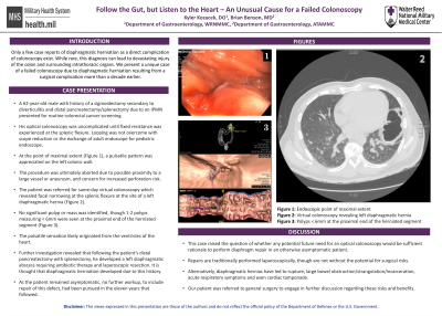
Case Description/Methods: A 62-year-old male with history of a sigmoidectomy secondary to diverticulitis and distal pancreatectomy/splenectomy due to an IPMN presented for routine colorectal cancer screening. His optical colonoscopy was uncomplicated until looping was experienced at the splenic flexure. Looping was not overcome with scope reduction or the exchange of adult endoscope for pediatric endoscope. At the point of maximal extent (Figure 1), a pulsatile pattern was appreciated on the left colonic wall. The procedure was ultimately aborted due to possible proximity to a large vessel or aneurysm, and concern for increased perforation risk.
The patient was referred for same-day virtual colonoscopy which revealed focal narrowing at the splenic flexure at the site of a left diaphragmatic hernia (Figure 2). No significant polyp or mass was identified, though 1-2 polyps measuring < 6mm were seen at the proximal end of the herniated segment (Figure 3). The pulsatile sensation likely originated from the ventricles of the heart.
Further investigation revealed that following the patient’s distal pancreatectomy with splenectomy, he developed a left diaphragmatic abscess requiring antibiotic therapy and laparoscopic resection. It is thought that diaphragmatic herniation developed due to this history. As the patient remained asymptomatic, no further workup, to include repair of this defect, had been pursued in the eleven years that followed.
Discussion: This case raised the question of whether any potential future need for an optical colonoscopy would be sufficient rationale to perform diaphragm repair in an otherwise asymptomatic patient. These repairs are traditionally performed laparoscopically, but are not without the potential for infection, adhesions and other surgical risks. On the other hand, diaphragmatic hernias have led to rupture, large bowel obstruction/strangulation/incarceration, acute respiratory symptoms and even cardiac tamponade. Our patient was referred to general surgery to engage in further discussion regarding these risks and benefits.

Disclosures:
Kyler Kozacek, DO1, Brian Benson, MD2. P3213 - Follow the Gut, but Listen to the Heart – An Unusual Cause for a Failed Colonoscopy, ACG 2023 Annual Scientific Meeting Abstracts. Vancouver, BC, Canada: American College of Gastroenterology.
1Walter Reed National Military Medical Center, Bethesda, MD; 2Fort Belvoir Community Hospital, Fort Belvoir, MD
Introduction: There are only a few case reports of diaphragmatic herniation as a direct complication of a colonoscopy. While rare, this diagnosis can lead to devastating injury of the colon and surrounding intrathoracic organs. We present a unique case of a failed colonoscopy due to diaphragmatic herniation resulting from a surgical complication more than a decade earlier.
Case Description/Methods: A 62-year-old male with history of a sigmoidectomy secondary to diverticulitis and distal pancreatectomy/splenectomy due to an IPMN presented for routine colorectal cancer screening. His optical colonoscopy was uncomplicated until looping was experienced at the splenic flexure. Looping was not overcome with scope reduction or the exchange of adult endoscope for pediatric endoscope. At the point of maximal extent (Figure 1), a pulsatile pattern was appreciated on the left colonic wall. The procedure was ultimately aborted due to possible proximity to a large vessel or aneurysm, and concern for increased perforation risk.
The patient was referred for same-day virtual colonoscopy which revealed focal narrowing at the splenic flexure at the site of a left diaphragmatic hernia (Figure 2). No significant polyp or mass was identified, though 1-2 polyps measuring < 6mm were seen at the proximal end of the herniated segment (Figure 3). The pulsatile sensation likely originated from the ventricles of the heart.
Further investigation revealed that following the patient’s distal pancreatectomy with splenectomy, he developed a left diaphragmatic abscess requiring antibiotic therapy and laparoscopic resection. It is thought that diaphragmatic herniation developed due to this history. As the patient remained asymptomatic, no further workup, to include repair of this defect, had been pursued in the eleven years that followed.
Discussion: This case raised the question of whether any potential future need for an optical colonoscopy would be sufficient rationale to perform diaphragm repair in an otherwise asymptomatic patient. These repairs are traditionally performed laparoscopically, but are not without the potential for infection, adhesions and other surgical risks. On the other hand, diaphragmatic hernias have led to rupture, large bowel obstruction/strangulation/incarceration, acute respiratory symptoms and even cardiac tamponade. Our patient was referred to general surgery to engage in further discussion regarding these risks and benefits.

Figure: Figure 1 - Endoscopic view of maximal extent
Figure 2 - Cross-sectional imaging of splenic flexure narrowing at left diaphragm hernia site
Figure 3 - Polyps measuring <6mm seen by virtual colonoscopy at the proximal end of the herniated segment
Figure 2 - Cross-sectional imaging of splenic flexure narrowing at left diaphragm hernia site
Figure 3 - Polyps measuring <6mm seen by virtual colonoscopy at the proximal end of the herniated segment
Disclosures:
Kyler Kozacek indicated no relevant financial relationships.
Brian Benson indicated no relevant financial relationships.
Kyler Kozacek, DO1, Brian Benson, MD2. P3213 - Follow the Gut, but Listen to the Heart – An Unusual Cause for a Failed Colonoscopy, ACG 2023 Annual Scientific Meeting Abstracts. Vancouver, BC, Canada: American College of Gastroenterology.
