Tuesday Poster Session
Category: Stomach
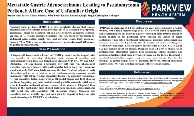
P4221 - Metastatic Gastric Adenocarcinoma Leading to Pseudomyxoma Peritonei: A Rare Case of Unfamiliar Origin
Tuesday, October 24, 2023
10:30 AM - 4:00 PM PT
Location: Exhibit Hall

Has Audio
.jpg)
Bryant Pinto Javier, DO, MHA
Parkview Medical Center
Pueblo, CO
Presenting Author(s)
Bryant Pinto. Javier, DO, MHA, Aleena Sammar, MD, Uday Patel, DO, Kumari Piryanka, MD, Rajiv Singh, MD, Christopher Calcagno, DO
Parkview Medical Center, Pueblo, CO
Introduction: Pseudomyxoma peritonei (PMP) is a rare neoplastic disease that causes mucinous ascites or deposits in the peritoneal cavity. It is caused by a ruptured appendiceal mucinous neoplasm but can also be rarely caused by ovarian, urachus, or GI-related cancers. Symptoms can vary from asymptomatic to abdominal pain, ascites, weight loss, and digestive issues. Early diagnosis through CT or MRI is crucial. We present a rare case of advanced PMP caused by gastric adenocarcinoma.
Case Description/Methods: A 66-year-old female with a history of GERD presented to the hospital with two months of worsening abdominal pain, decreased appetite, and unintentional weight loss. Lab tests showed elevated ALP, CA 19-9, and CEA. Abdominal CT scan showed a lobulated liver with thin rim enhancement extending into porta hepatis with small nodular foci in the greater omentum consistent with PMP, completely encasing the liver. Evidence of gastric body thickening and periaortic and pericaval lymphadenopathy suggested gastric malignancy with intraperitoneal metastatic disease. The appendix was normal. GI was consulted. Patient underwent an IR-guided biopsy and EGD showing an 8 cm fungating, polypoid, and ulcerated partially circumferential mass. Gastric biopsies confirmed adenocarcinoma of the stomach. CT-guided liver biopsy in the perihepatic mass showed metastatic mucinous adenocarcinoma with signet ring cells consistent with metastatic disease. Oncology was consulted, and a chemotherapy port with plan for outpatient follow up with surgical oncology for PET/CT and debulking.
Discussion: PMP has an incidence of 1-2 per million per year, more commonly affecting women, with a mean incidence age of 53. PMP is often found in appendiceal and ovarian tumors but rarely in digestive system tumors. PMP is caused by tumor rupture in the peritoneal cavity causing the spread of mucin-containing tumor cells or peritoneal metastasis of mucinous adenocarcinoma. Copious mucinous fluid gradually fills the peritoneal cavity, resulting in a “jelly belly” abdomen. Elevated tumor markers such as CEA, CA 19-9, and CA 125 indicate advanced disease. Diagnosis with CT or MRI shows low or proteinaceous attenuation ascites, liver scalloping, splenic margins, and peritoneal implants that cause extrinsic pressure on the bowel. Treatment is maximal cytoreduction surgery completed and chemotherapy. Few data for survival in gastric-origin PMP is available. However, without treatment, gastric-origin PMP has a median survival of three to four months.
Disclosures:
Bryant Pinto. Javier, DO, MHA, Aleena Sammar, MD, Uday Patel, DO, Kumari Piryanka, MD, Rajiv Singh, MD, Christopher Calcagno, DO. P4221 - Metastatic Gastric Adenocarcinoma Leading to Pseudomyxoma Peritonei: A Rare Case of Unfamiliar Origin, ACG 2023 Annual Scientific Meeting Abstracts. Vancouver, BC, Canada: American College of Gastroenterology.
Parkview Medical Center, Pueblo, CO
Introduction: Pseudomyxoma peritonei (PMP) is a rare neoplastic disease that causes mucinous ascites or deposits in the peritoneal cavity. It is caused by a ruptured appendiceal mucinous neoplasm but can also be rarely caused by ovarian, urachus, or GI-related cancers. Symptoms can vary from asymptomatic to abdominal pain, ascites, weight loss, and digestive issues. Early diagnosis through CT or MRI is crucial. We present a rare case of advanced PMP caused by gastric adenocarcinoma.
Case Description/Methods: A 66-year-old female with a history of GERD presented to the hospital with two months of worsening abdominal pain, decreased appetite, and unintentional weight loss. Lab tests showed elevated ALP, CA 19-9, and CEA. Abdominal CT scan showed a lobulated liver with thin rim enhancement extending into porta hepatis with small nodular foci in the greater omentum consistent with PMP, completely encasing the liver. Evidence of gastric body thickening and periaortic and pericaval lymphadenopathy suggested gastric malignancy with intraperitoneal metastatic disease. The appendix was normal. GI was consulted. Patient underwent an IR-guided biopsy and EGD showing an 8 cm fungating, polypoid, and ulcerated partially circumferential mass. Gastric biopsies confirmed adenocarcinoma of the stomach. CT-guided liver biopsy in the perihepatic mass showed metastatic mucinous adenocarcinoma with signet ring cells consistent with metastatic disease. Oncology was consulted, and a chemotherapy port with plan for outpatient follow up with surgical oncology for PET/CT and debulking.
Discussion: PMP has an incidence of 1-2 per million per year, more commonly affecting women, with a mean incidence age of 53. PMP is often found in appendiceal and ovarian tumors but rarely in digestive system tumors. PMP is caused by tumor rupture in the peritoneal cavity causing the spread of mucin-containing tumor cells or peritoneal metastasis of mucinous adenocarcinoma. Copious mucinous fluid gradually fills the peritoneal cavity, resulting in a “jelly belly” abdomen. Elevated tumor markers such as CEA, CA 19-9, and CA 125 indicate advanced disease. Diagnosis with CT or MRI shows low or proteinaceous attenuation ascites, liver scalloping, splenic margins, and peritoneal implants that cause extrinsic pressure on the bowel. Treatment is maximal cytoreduction surgery completed and chemotherapy. Few data for survival in gastric-origin PMP is available. However, without treatment, gastric-origin PMP has a median survival of three to four months.
Disclosures:
Bryant Javier indicated no relevant financial relationships.
Aleena Sammar indicated no relevant financial relationships.
Uday Patel indicated no relevant financial relationships.
Kumari Piryanka indicated no relevant financial relationships.
Rajiv Singh indicated no relevant financial relationships.
Christopher Calcagno indicated no relevant financial relationships.
Bryant Pinto. Javier, DO, MHA, Aleena Sammar, MD, Uday Patel, DO, Kumari Piryanka, MD, Rajiv Singh, MD, Christopher Calcagno, DO. P4221 - Metastatic Gastric Adenocarcinoma Leading to Pseudomyxoma Peritonei: A Rare Case of Unfamiliar Origin, ACG 2023 Annual Scientific Meeting Abstracts. Vancouver, BC, Canada: American College of Gastroenterology.
