Tuesday Poster Session
Category: Small Intestine
P4119 - Small Bowel Lymphoma: A Case Series
Tuesday, October 24, 2023
10:30 AM - 4:00 PM PT
Location: Exhibit Hall

Has Audio

Sharon Slomovich, MD
Mount Sinai South Nassau
Oceanside, NY
Presenting Author(s)
Sharon Slomovich, MD1, Mahnoor Khan, DO2, Neal Shah, DO1, Frank Gress, MD, MBA, FACG3
1Mount Sinai South Nassau, Oceanside, NY; 2ISMMS Mt. Sinai South Nassau, Oceanside, NY; 3Icahn School of Medicine at Mount Sinai (ISMMS), Mount Sinai South Nassau (MSSN), Oceanside, NY
Introduction: Lymphoma originating from the gastrointestinal tract is rare and accounts for only 1-4% of malignancies arising in the stomach, small intestine or colon. Patients often present with nonspecific symptoms and despite the application of advanced imaging techniques, diagnosis can be challenging. We present a case series of two patients with ambiguous gastrointestinal symptoms and a subsequent diagnosis of primary intestinal lymphoma.
Case Description/Methods: A 43-year-old male with a medical history of sarcoidosis presented with three months of periumbilical pain and a 30-pound, unintentional weight loss. Physical exam, laboratory investigation, esophagogastroduodenoscopy (EGD), colonoscopy and video capsule endoscopy (VCE) were unremarkable. CT abdomen and pelvis revealed a partial small bowel obstruction (Figure 1a). A subsequent small bowel enteroscopy (SBE) demonstrated a nonspecific inflammation of the ileum. Additionally, a CT enterography disclosed the presence of a stricture in the mid-ileum. Given the unclear etiology of the pathology three months following presentation, small bowel resection was performed and pathology revealed a diffuse large B-cell lymphoma.
A 55-year-old female with a medical history of Lynch syndrome presented with one month of abdominal pain, vomiting and a five-pound, unintentional weight loss. The physical exam and laboratory investigation were normal. Magnetic resonance enterography showed a 6 cm proximal ileal segment with irregular concentric wall thickening. SBE revealed nodular ileal mucosa with ulceration in the mid-ileum (Figure 1b). Biopsies demonstrated low-grade follicular lymphoma and this was demonstrated four months after presentation.
Discussion: Presenting symptoms of primary intestinal lymphoma are vague and commonly include abdominal pain, hematochezia, melena or changes in bowel habits. Radiological findings are often nonspecific and may present a challenge in distinguishing lymphoma from other lesions, benign or malignant. While VCE and small bowel enteroscopy with biopsies have improved the identification of small intestinal pathologies, a nonspecific clinical presentation can result in delayed intervention. Thus, diagnosing primary intestinal lymphoma remains a challenge and further research is needed to provide insight on the best practice for its diagnosis and treatment.

Disclosures:
Sharon Slomovich, MD1, Mahnoor Khan, DO2, Neal Shah, DO1, Frank Gress, MD, MBA, FACG3. P4119 - Small Bowel Lymphoma: A Case Series, ACG 2023 Annual Scientific Meeting Abstracts. Vancouver, BC, Canada: American College of Gastroenterology.
1Mount Sinai South Nassau, Oceanside, NY; 2ISMMS Mt. Sinai South Nassau, Oceanside, NY; 3Icahn School of Medicine at Mount Sinai (ISMMS), Mount Sinai South Nassau (MSSN), Oceanside, NY
Introduction: Lymphoma originating from the gastrointestinal tract is rare and accounts for only 1-4% of malignancies arising in the stomach, small intestine or colon. Patients often present with nonspecific symptoms and despite the application of advanced imaging techniques, diagnosis can be challenging. We present a case series of two patients with ambiguous gastrointestinal symptoms and a subsequent diagnosis of primary intestinal lymphoma.
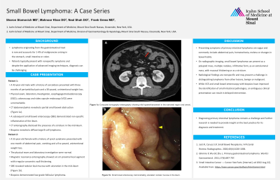
Case Description/Methods: A 43-year-old male with a medical history of sarcoidosis presented with three months of periumbilical pain and a 30-pound, unintentional weight loss. Physical exam, laboratory investigation, esophagogastroduodenoscopy (EGD), colonoscopy and video capsule endoscopy (VCE) were unremarkable. CT abdomen and pelvis revealed a partial small bowel obstruction (Figure 1a). A subsequent small bowel enteroscopy (SBE) demonstrated a nonspecific inflammation of the ileum. Additionally, a CT enterography disclosed the presence of a stricture in the mid-ileum. Given the unclear etiology of the pathology three months following presentation, small bowel resection was performed and pathology revealed a diffuse large B-cell lymphoma.
A 55-year-old female with a medical history of Lynch syndrome presented with one month of abdominal pain, vomiting and a five-pound, unintentional weight loss. The physical exam and laboratory investigation were normal. Magnetic resonance enterography showed a 6 cm proximal ileal segment with irregular concentric wall thickening. SBE revealed nodular ileal mucosa with ulceration in the mid-ileum (Figure 1b). Biopsies demonstrated low-grade follicular lymphoma and this was demonstrated four months after presentation.
Discussion: Presenting symptoms of primary intestinal lymphoma are vague and commonly include abdominal pain, hematochezia, melena or changes in bowel habits. Radiological findings are often nonspecific and may present a challenge in distinguishing lymphoma from other lesions, benign or malignant. While VCE and small bowel enteroscopy with biopsies have improved the identification of small intestinal pathologies, a nonspecific clinical presentation can result in delayed intervention. Thus, diagnosing primary intestinal lymphoma remains a challenge and further research is needed to provide insight on the best practice for its diagnosis and treatment.

Figure: Figure 1a: Computed tomography enterography showing mild hyperenhancement in the narrowed region (red arrow). Figure 1b: Small bowel enteroscopy demonstrating ulcerated nodular mucosa in the ileum.
Disclosures:
Sharon Slomovich indicated no relevant financial relationships.
Mahnoor Khan indicated no relevant financial relationships.
Neal Shah indicated no relevant financial relationships.
Frank Gress: Salvo Health – Advisory Committee/Board Member, Stock Options.
Sharon Slomovich, MD1, Mahnoor Khan, DO2, Neal Shah, DO1, Frank Gress, MD, MBA, FACG3. P4119 - Small Bowel Lymphoma: A Case Series, ACG 2023 Annual Scientific Meeting Abstracts. Vancouver, BC, Canada: American College of Gastroenterology.
