Tuesday Poster Session
Category: Colon
P3123 - CLL: An Important Differential in the Case of Microscopic Colitis
Tuesday, October 24, 2023
10:30 AM - 4:00 PM PT
Location: Exhibit Hall

Has Audio

Adil Ghafoor, MD
Advocate Aurora Health
Brookfield, Wisconsin
Presenting Author(s)
Adil Ghafoor, MD1, Nadia Huq, MD2
1Advocate Aurora Health, Brookfield, WI; 2Advocate Aurora Health, Milwaukee, WI
Introduction: Chronic lymphocytic leukemia/small lymphocytic lymphoma (CLL/SLL) typically presents as asymptomatic lymphocytosis or leukocytosis and/or in the setting of lymphadenopathy, recurrent infections, or other hematologic abnormalities. GI involvement is a rare occurrence and can be associated with the aggressive diffuse large B-cell lymphoma or Richter’s transformation. We outline a case of colonic involvement of CLL/SLL found on diagnostic colonoscopy for diarrhea in a patient with a history of CLL/SLL previously in remission.
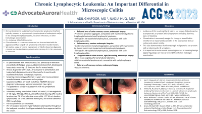
Case Description/Methods: A 61-year-old male with a history of CLL/SLL previously in remission presented with fatigue, urgency, abdominal discomfort, bloating and loose stools occurring 1-2 times per day for several weeks. He was diagnosed with CLL/SLL 6 years prior to presentation and treated with Bendamustine and Rituximab for 5 months with excellent clinical and hematologic response. Screening colonoscopy performed 11 years prior to presentation revealed internal hemorrhoids and no polyps. Supplemental fiber, a 4-week trial of low FODMAP diet and diagnostic colonoscopy were recommended. Colonoscopy revealed one cecal polyp, one ascending colon polyp, one rectal polyp, and internal hemorrhoids. Cecal polyp biopsy revealed prominent lymphoid aggregate compatible with CLL/SLL and mild intraepithelial lymphocytosis compatible with early lymphocytic colitis. Ascending colon polyp biopsy revealed a tubular adenoma with similar findings of mild intraepithelial lymphocytosis compatible with early lymphocytic colitis. Random colon biopsies revealed scattered prominent lymphoid aggregates also compatible with CLL/SLL. The patient was trialed on budesonide with no symptomatic improvement. Laboratory testing revealed an LDH of 249 units/L, B2 microglobulin of 4.0 mg/L, WBC 22.2 K/mcL, with differential showing 14% reactive lymphocytes, 7.8 K/mcL absolute neutrophils, 12.7 K/mcL absolute lymphocytes, 1.8 K/mcL absolute monocytes, and overall abnormal WBC morphology. Fish CLL panel was unremarkable. PET CT revealed extensive hypermetabolic adenopathy throughout the body and a modest sized hypermetabolic focus apparent within the cecum.
Discussion: GI evaluation is commonly sought for change in bowel habits. It is important to consider, in the appropriate clinical setting, hematologic malignancies can present with predominantly GI symptoms and benign appearing polyps can have a varied differential on histologic examination.
Disclosures:
Adil Ghafoor, MD1, Nadia Huq, MD2. P3123 - CLL: An Important Differential in the Case of Microscopic Colitis, ACG 2023 Annual Scientific Meeting Abstracts. Vancouver, BC, Canada: American College of Gastroenterology.
1Advocate Aurora Health, Brookfield, WI; 2Advocate Aurora Health, Milwaukee, WI
Introduction: Chronic lymphocytic leukemia/small lymphocytic lymphoma (CLL/SLL) typically presents as asymptomatic lymphocytosis or leukocytosis and/or in the setting of lymphadenopathy, recurrent infections, or other hematologic abnormalities. GI involvement is a rare occurrence and can be associated with the aggressive diffuse large B-cell lymphoma or Richter’s transformation. We outline a case of colonic involvement of CLL/SLL found on diagnostic colonoscopy for diarrhea in a patient with a history of CLL/SLL previously in remission.
Case Description/Methods: A 61-year-old male with a history of CLL/SLL previously in remission presented with fatigue, urgency, abdominal discomfort, bloating and loose stools occurring 1-2 times per day for several weeks. He was diagnosed with CLL/SLL 6 years prior to presentation and treated with Bendamustine and Rituximab for 5 months with excellent clinical and hematologic response. Screening colonoscopy performed 11 years prior to presentation revealed internal hemorrhoids and no polyps. Supplemental fiber, a 4-week trial of low FODMAP diet and diagnostic colonoscopy were recommended. Colonoscopy revealed one cecal polyp, one ascending colon polyp, one rectal polyp, and internal hemorrhoids. Cecal polyp biopsy revealed prominent lymphoid aggregate compatible with CLL/SLL and mild intraepithelial lymphocytosis compatible with early lymphocytic colitis. Ascending colon polyp biopsy revealed a tubular adenoma with similar findings of mild intraepithelial lymphocytosis compatible with early lymphocytic colitis. Random colon biopsies revealed scattered prominent lymphoid aggregates also compatible with CLL/SLL. The patient was trialed on budesonide with no symptomatic improvement. Laboratory testing revealed an LDH of 249 units/L, B2 microglobulin of 4.0 mg/L, WBC 22.2 K/mcL, with differential showing 14% reactive lymphocytes, 7.8 K/mcL absolute neutrophils, 12.7 K/mcL absolute lymphocytes, 1.8 K/mcL absolute monocytes, and overall abnormal WBC morphology. Fish CLL panel was unremarkable. PET CT revealed extensive hypermetabolic adenopathy throughout the body and a modest sized hypermetabolic focus apparent within the cecum.
Discussion: GI evaluation is commonly sought for change in bowel habits. It is important to consider, in the appropriate clinical setting, hematologic malignancies can present with predominantly GI symptoms and benign appearing polyps can have a varied differential on histologic examination.
Disclosures:
Adil Ghafoor indicated no relevant financial relationships.
Nadia Huq indicated no relevant financial relationships.
Adil Ghafoor, MD1, Nadia Huq, MD2. P3123 - CLL: An Important Differential in the Case of Microscopic Colitis, ACG 2023 Annual Scientific Meeting Abstracts. Vancouver, BC, Canada: American College of Gastroenterology.
