Tuesday Poster Session
Category: Colon
P3124 - A Case of Acute Diverticulitis Without Diverticula
Tuesday, October 24, 2023
10:30 AM - 4:00 PM PT
Location: Exhibit Hall

Has Audio

Patricia Sobczyk, BS
Creighton University School of Medicine
Omaha, NE
Presenting Author(s)
Patricia Sobczyk, BS1, Suhail Sidhu, BS1, Paul Millner, MD1, Saurabh Chandan, MD, MBBS1, Sandeep Mukherjee, MD2
1Creighton University School of Medicine, Omaha, NE; 2Creighton University, Omaha, NE
Introduction: Acute diverticulitis is inflammation of pouches in the colon (diverticula) that occurs in 4% of
patients with diverticulosis. Follow-up colonoscopy is recommended for patients that
experience diverticulitis to rule out malignancy, but the recommendation is under scrutiny. Most
sensitive findings on CT Scan include bowel wall thickening and fat stranding; most specific
findings include abscesses, inflamed diverticulum, intramural air, and phlegmon. We describe a
case of acute diverticulitis diagnosed using physical exam, labs, and imaging but follow-up
colonoscopy did not show diverticula.
Case Description/Methods: A 38-year-old female with past medical history of migraines and a past surgical history of left
ovarian embolization with left oophorectomy dilation and curettage presented with left lower
quadrant pain for three days which migrated to the suprapubic region in the right lower quadrant.
She denied bleeding, fever, nausea, and vomiting. Physical exam was pertinent for tenderness of
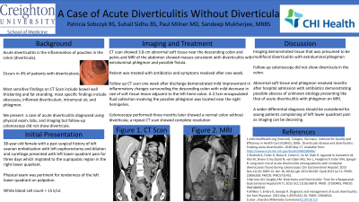
the left lower quadrant on palpation. White blood cell count was 15 k/ul. CT scan showed 3.8cm
abnormal soft tissue near the descending colon and pelvis. Gynecology was consulted and did
not deem it consistent with a gynecological etiology. MRI of the abdomen showed masses
consistent with diverticulitis with extraluminal phlegmon and possible fistula. Patient was treated
with antibiotics and symptoms resolved after one week. Follow up CT scan one week after
discharge demonstrated mild improvement in inflammatory changes surrounding the descending
colon with mild decrease in size of soft tissue lesion adjacent to the left hemicolon. A 2.5cm
encapsulated fluid collection involving the possible phlegmon was located near the right
hemipelvis; interventional radiology denied needing drainage. Colonoscopy performed three
months later showed a normal colon without diverticula; a repeat CT scan was normal.
Discussion: In this young patient, imaging demonstrated tissue that was presumed to be multifocal
diverticulitis with extraluminal phlegmon. However, follow-up colonoscopy did not show
diverticula in the colon. The abnormal soft tissue and phlegmon resolved months after hospital
admission with antibiotics demonstrating possible abscess of unknown etiology presenting
similar to that of acute diverticulitis with phlegmon on MRI. A wider differential diagnosis
should be considered for young patients complaining of left lower quadrant pain with elevated
white blood cell counts as imaging can be deceiving.

Disclosures:
Patricia Sobczyk, BS1, Suhail Sidhu, BS1, Paul Millner, MD1, Saurabh Chandan, MD, MBBS1, Sandeep Mukherjee, MD2. P3124 - A Case of Acute Diverticulitis Without Diverticula, ACG 2023 Annual Scientific Meeting Abstracts. Vancouver, BC, Canada: American College of Gastroenterology.
1Creighton University School of Medicine, Omaha, NE; 2Creighton University, Omaha, NE
Introduction: Acute diverticulitis is inflammation of pouches in the colon (diverticula) that occurs in 4% of
patients with diverticulosis. Follow-up colonoscopy is recommended for patients that
experience diverticulitis to rule out malignancy, but the recommendation is under scrutiny. Most
sensitive findings on CT Scan include bowel wall thickening and fat stranding; most specific
findings include abscesses, inflamed diverticulum, intramural air, and phlegmon. We describe a
case of acute diverticulitis diagnosed using physical exam, labs, and imaging but follow-up
colonoscopy did not show diverticula.
Case Description/Methods: A 38-year-old female with past medical history of migraines and a past surgical history of left
ovarian embolization with left oophorectomy dilation and curettage presented with left lower
quadrant pain for three days which migrated to the suprapubic region in the right lower quadrant.
She denied bleeding, fever, nausea, and vomiting. Physical exam was pertinent for tenderness of
the left lower quadrant on palpation. White blood cell count was 15 k/ul. CT scan showed 3.8cm
abnormal soft tissue near the descending colon and pelvis. Gynecology was consulted and did
not deem it consistent with a gynecological etiology. MRI of the abdomen showed masses
consistent with diverticulitis with extraluminal phlegmon and possible fistula. Patient was treated
with antibiotics and symptoms resolved after one week. Follow up CT scan one week after
discharge demonstrated mild improvement in inflammatory changes surrounding the descending
colon with mild decrease in size of soft tissue lesion adjacent to the left hemicolon. A 2.5cm
encapsulated fluid collection involving the possible phlegmon was located near the right
hemipelvis; interventional radiology denied needing drainage. Colonoscopy performed three
months later showed a normal colon without diverticula; a repeat CT scan was normal.
Discussion: In this young patient, imaging demonstrated tissue that was presumed to be multifocal
diverticulitis with extraluminal phlegmon. However, follow-up colonoscopy did not show
diverticula in the colon. The abnormal soft tissue and phlegmon resolved months after hospital
admission with antibiotics demonstrating possible abscess of unknown etiology presenting
similar to that of acute diverticulitis with phlegmon on MRI. A wider differential diagnosis
should be considered for young patients complaining of left lower quadrant pain with elevated
white blood cell counts as imaging can be deceiving.

Figure: Figure 1: Masses adjacent to the left colon and within the anatomic pelvis. Favor multifocal diverticulitis with extraluminal phlegmon and possibly developing fistulas. Disease likely involves the right ovary.
Disclosures:
Patricia Sobczyk indicated no relevant financial relationships.
Suhail Sidhu indicated no relevant financial relationships.
Paul Millner indicated no relevant financial relationships.
Saurabh Chandan indicated no relevant financial relationships.
Sandeep Mukherjee: Dynamed Plus – Section editor for Hepatology. Gilead – Speakers Bureau.
Patricia Sobczyk, BS1, Suhail Sidhu, BS1, Paul Millner, MD1, Saurabh Chandan, MD, MBBS1, Sandeep Mukherjee, MD2. P3124 - A Case of Acute Diverticulitis Without Diverticula, ACG 2023 Annual Scientific Meeting Abstracts. Vancouver, BC, Canada: American College of Gastroenterology.
