Tuesday Poster Session
Category: Small Intestine
P4125 - Symptomatic Jejunal Diverticulosis: A Rare Entity and a Cause of Recurrent Small Bowel Obstruction
Tuesday, October 24, 2023
10:30 AM - 4:00 PM PT
Location: Exhibit Hall

Has Audio

Vatsal Khanna, MD
Wayne State University School of Medicine
Rochester Hills, MI
Presenting Author(s)
Vatsal Khanna, MD1, Trishya Reddy, MD1, Eric Martin Sieloff, MD2, Tripti Nagar, MD1, Pranav Chalasani, MD1
1Wayne State University School of Medicine, Rochester Hills, MI; 2Ascension Providence Southfield Hospital/ Michigan State University, Southfield, MI
Introduction: Jejunal diverticular disease is a rare entity with a higher prevalence after the 6th decade of life. It is usually asymptomatic, and patients typically present with vague symptoms like malabsorption, pain, or nausea. Complications are rare and occur only in 10% of patients. We report a unique case of a 71-year-old male who presented with a history of chronic diarrhea and recurrent attacks of partial small bowel obstruction and was found to have a large jejunal diverticulum during diagnostic laparoscopy and subsequently underwent small bowel resection and anastomosis.
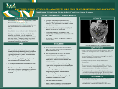
Case Description/Methods: A 71-year-old male with a history of coronary artery disease, unintentional weight loss, chronic diarrhea, and recurrent episodes of partial small bowel obstruction (SBO) was admitted to our hospital with complaints of diffuse abdominal pain and nausea. CT (computed tomography) Enterography done in the past revealed multiple jejunal diverticulosis. On admission, the patient was hemodynamically stable and afebrile. Clinical laboratory results were unremarkable. Physical examination revealed mild diffuse abdominal tenderness with no signs of peritonitis. General Surgery was consulted. The patient had a diagnostic laparoscopy which showed a large jejunal diverticulum proximal to the ligament of Treitz measuring 8 cm, along with multiple small jejunal diverticula. He subsequently underwent segmental small bowel resection and end-to-end anastomosis. The postoperative period was uneventful, and histopathology revealed prominent diverticulosis of the small bowel. On follow up, the patient was symptom free with no episodes of SBO.
Discussion: Jejunal diverticula are rare, with a clinical incidence ranging from 0.06% to 1.3% and a higher prevalence after the 6th decade of life. They are predominantly localized to the proximal jejunum, followed by the distal jejunum. It is believed to arise from abnormalities in the smooth muscle intestinal dyskinesia, and high intra-luminal pressures. Due to nonspecific clinical presentation, the diagnosis is often difficult and delayed, with patients presenting with vague abdominal pain, nausea, or malabsorption. In addition, 10 to 30% of patients can develop acute complications like infection, bowel perforation, volvulus, or bleeding. CT abdomen with contrast is the investigation of choice due to high diagnostic accuracy. Treatment is usually conservative. Surgery is reserved for patients with complications and involves resection and primary anastomosis.

Disclosures:
Vatsal Khanna, MD1, Trishya Reddy, MD1, Eric Martin Sieloff, MD2, Tripti Nagar, MD1, Pranav Chalasani, MD1. P4125 - Symptomatic Jejunal Diverticulosis: A Rare Entity and a Cause of Recurrent Small Bowel Obstruction, ACG 2023 Annual Scientific Meeting Abstracts. Vancouver, BC, Canada: American College of Gastroenterology.
1Wayne State University School of Medicine, Rochester Hills, MI; 2Ascension Providence Southfield Hospital/ Michigan State University, Southfield, MI
Introduction: Jejunal diverticular disease is a rare entity with a higher prevalence after the 6th decade of life. It is usually asymptomatic, and patients typically present with vague symptoms like malabsorption, pain, or nausea. Complications are rare and occur only in 10% of patients. We report a unique case of a 71-year-old male who presented with a history of chronic diarrhea and recurrent attacks of partial small bowel obstruction and was found to have a large jejunal diverticulum during diagnostic laparoscopy and subsequently underwent small bowel resection and anastomosis.
Case Description/Methods: A 71-year-old male with a history of coronary artery disease, unintentional weight loss, chronic diarrhea, and recurrent episodes of partial small bowel obstruction (SBO) was admitted to our hospital with complaints of diffuse abdominal pain and nausea. CT (computed tomography) Enterography done in the past revealed multiple jejunal diverticulosis. On admission, the patient was hemodynamically stable and afebrile. Clinical laboratory results were unremarkable. Physical examination revealed mild diffuse abdominal tenderness with no signs of peritonitis. General Surgery was consulted. The patient had a diagnostic laparoscopy which showed a large jejunal diverticulum proximal to the ligament of Treitz measuring 8 cm, along with multiple small jejunal diverticula. He subsequently underwent segmental small bowel resection and end-to-end anastomosis. The postoperative period was uneventful, and histopathology revealed prominent diverticulosis of the small bowel. On follow up, the patient was symptom free with no episodes of SBO.
Discussion: Jejunal diverticula are rare, with a clinical incidence ranging from 0.06% to 1.3% and a higher prevalence after the 6th decade of life. They are predominantly localized to the proximal jejunum, followed by the distal jejunum. It is believed to arise from abnormalities in the smooth muscle intestinal dyskinesia, and high intra-luminal pressures. Due to nonspecific clinical presentation, the diagnosis is often difficult and delayed, with patients presenting with vague abdominal pain, nausea, or malabsorption. In addition, 10 to 30% of patients can develop acute complications like infection, bowel perforation, volvulus, or bleeding. CT abdomen with contrast is the investigation of choice due to high diagnostic accuracy. Treatment is usually conservative. Surgery is reserved for patients with complications and involves resection and primary anastomosis.

Figure: CT Enterography demonstrating jejunal diverticulosis
Disclosures:
Vatsal Khanna indicated no relevant financial relationships.
Trishya Reddy indicated no relevant financial relationships.
Eric Martin Sieloff indicated no relevant financial relationships.
Tripti Nagar indicated no relevant financial relationships.
Pranav Chalasani indicated no relevant financial relationships.
Vatsal Khanna, MD1, Trishya Reddy, MD1, Eric Martin Sieloff, MD2, Tripti Nagar, MD1, Pranav Chalasani, MD1. P4125 - Symptomatic Jejunal Diverticulosis: A Rare Entity and a Cause of Recurrent Small Bowel Obstruction, ACG 2023 Annual Scientific Meeting Abstracts. Vancouver, BC, Canada: American College of Gastroenterology.
