Tuesday Poster Session
Category: Liver
P3898 - Spindle Cell Carcinoma: An Exceedingly Rare Liver Biopsy Proven Case
Tuesday, October 24, 2023
10:30 AM - 4:00 PM PT
Location: Exhibit Hall

Has Audio

Sanya Goswami, MD
SUNY Downstate Health Sciences University
Brooklyn, NY
Presenting Author(s)
Sanya Goswami, MD1, Qi Yu, MD, MPH2, Binyaman R. Abramowitz, MD1, Fazl Rahim Wazeen, MD3
1SUNY Downstate Health Sciences University, Brooklyn, NY; 2SUNY Downstate Health Sciences University/NYCHHC Kings County Hospital, Brooklyn, NY; 3Greater Baltimore Medical Center, Towson, MD
Introduction: Spindle cell neoplasms, also known as sarcomatoid carcinoma, are typically biphasic tumors, composed of conventional squamous cell carcinoma and malignant spindle cells. It occurs in primarily in the fifth or sixth decades of life, affects men, and can occur in soft tissue, bone, or any part of the body. Due to its rarity and lack of diagnostic schema in the literature, it is often an overlooked, and missed diagnosis.
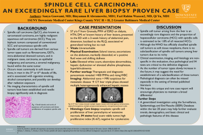
Case Description/Methods: Here, we highlight a rare case of a 57-year-old female from Grenada with PMH of ESRD on dialysis, HTN, and DM, with a known history of liver lesions, who presented to the ED with a 2-week history of abdominal pain and distension, localized to the right lower quadrant, associated with generalized itching, but no identifiable rash. Vitals on presentation were unremarkable and physical examination was remarkable for scleral icterus, excoriations on the abdomen, markedly distended, non-tender abdomen with a positive fluid wave and shifting dullness. Labs were remarkable for elevated white count, electrolyte abnormalities, elevated alkaline phosphatase, and hyperbilirubinemia. Diagnostic and therapeutic paracentesis revealed >400 PMNs and rising WBC. After abdominal sonogram and MRI revealed suspicion for metastatic disease, CT liver triple phase revealed multiple hepatic masses with heterogenous enhancement, enlarged surround lymph nodes, and CT chest showed multiple lung nodules. Pathology report of the liver biopsy revealed neoplastic spindle cell proliferation involving liver parenchyma and extensive necrosis. IH stains showed focal scant viable tumor and a high proliferative index (Ki-67).
Discussion: Spindle cell tumor arising from the liver is exceedingly rare and the proportion of hepatocellular carcinoma (HCC) with spindle cells is estimated to be 1.8% of all resected HCCs. There is no subclassification of spindle cell tumors among liver tumors. Pathological and IH tests are critical to the definitive diagnosis. As the number of tumor types slowly increase in the future, it may suggest the need for establishment of a subclassification of these tumors. We hope this unique and rare case report will encourage physicians to maintain a broad differential. Next steps could be a generalized investigation using the Surveillance, Epidemiology, and End Results (SEER) database within the last 20 years to help further evaluate trends, demographics, and basic clinical and pathologic features of this disease.

Disclosures:
Sanya Goswami, MD1, Qi Yu, MD, MPH2, Binyaman R. Abramowitz, MD1, Fazl Rahim Wazeen, MD3. P3898 - Spindle Cell Carcinoma: An Exceedingly Rare Liver Biopsy Proven Case, ACG 2023 Annual Scientific Meeting Abstracts. Vancouver, BC, Canada: American College of Gastroenterology.
1SUNY Downstate Health Sciences University, Brooklyn, NY; 2SUNY Downstate Health Sciences University/NYCHHC Kings County Hospital, Brooklyn, NY; 3Greater Baltimore Medical Center, Towson, MD
Introduction: Spindle cell neoplasms, also known as sarcomatoid carcinoma, are typically biphasic tumors, composed of conventional squamous cell carcinoma and malignant spindle cells. It occurs in primarily in the fifth or sixth decades of life, affects men, and can occur in soft tissue, bone, or any part of the body. Due to its rarity and lack of diagnostic schema in the literature, it is often an overlooked, and missed diagnosis.
Case Description/Methods: Here, we highlight a rare case of a 57-year-old female from Grenada with PMH of ESRD on dialysis, HTN, and DM, with a known history of liver lesions, who presented to the ED with a 2-week history of abdominal pain and distension, localized to the right lower quadrant, associated with generalized itching, but no identifiable rash. Vitals on presentation were unremarkable and physical examination was remarkable for scleral icterus, excoriations on the abdomen, markedly distended, non-tender abdomen with a positive fluid wave and shifting dullness. Labs were remarkable for elevated white count, electrolyte abnormalities, elevated alkaline phosphatase, and hyperbilirubinemia. Diagnostic and therapeutic paracentesis revealed >400 PMNs and rising WBC. After abdominal sonogram and MRI revealed suspicion for metastatic disease, CT liver triple phase revealed multiple hepatic masses with heterogenous enhancement, enlarged surround lymph nodes, and CT chest showed multiple lung nodules. Pathology report of the liver biopsy revealed neoplastic spindle cell proliferation involving liver parenchyma and extensive necrosis. IH stains showed focal scant viable tumor and a high proliferative index (Ki-67).
Discussion: Spindle cell tumor arising from the liver is exceedingly rare and the proportion of hepatocellular carcinoma (HCC) with spindle cells is estimated to be 1.8% of all resected HCCs. There is no subclassification of spindle cell tumors among liver tumors. Pathological and IH tests are critical to the definitive diagnosis. As the number of tumor types slowly increase in the future, it may suggest the need for establishment of a subclassification of these tumors. We hope this unique and rare case report will encourage physicians to maintain a broad differential. Next steps could be a generalized investigation using the Surveillance, Epidemiology, and End Results (SEER) database within the last 20 years to help further evaluate trends, demographics, and basic clinical and pathologic features of this disease.

Figure: Figure 1.a and 1.b: CT liver 3 phase showed multiple hepatic masses with heterogenous enhancement,
atypical hemangiomas, multiple peripancreatic/paraaortic, retroperitoneal lymph nodes
atypical hemangiomas, multiple peripancreatic/paraaortic, retroperitoneal lymph nodes
Disclosures:
Sanya Goswami indicated no relevant financial relationships.
Qi Yu indicated no relevant financial relationships.
Binyaman Abramowitz indicated no relevant financial relationships.
Fazl Rahim Wazeen indicated no relevant financial relationships.
Sanya Goswami, MD1, Qi Yu, MD, MPH2, Binyaman R. Abramowitz, MD1, Fazl Rahim Wazeen, MD3. P3898 - Spindle Cell Carcinoma: An Exceedingly Rare Liver Biopsy Proven Case, ACG 2023 Annual Scientific Meeting Abstracts. Vancouver, BC, Canada: American College of Gastroenterology.
