Tuesday Poster Session
Category: Biliary/Pancreas
P2898 - Pancreaticopleural Fistula: An Uncommon Complication of Chronic Pancreatitis
Tuesday, October 24, 2023
10:30 AM - 4:00 PM PT
Location: Exhibit Hall

Has Audio

Gina Moon, MD
University of Oklahoma Health Sciences Center
Oklahoma City, OK
Presenting Author(s)
Gina Moon, MD1, Ahmad Basil Nasir, MD2, Muhammad Taha Khan, MD3, Tokunbo Ajayi, MD2, Amir Rumman, MD1
1University of Oklahoma Health Sciences Center, Oklahoma City, OK; 2University of Oklahoma College of Medicine, Oklahoma City, OK; 3Oklahoma University Health Sciences Center, Oklahoma City, OK
Introduction: Pancreaticopleural fistulas (PPFs) are usually seen in middle-aged men with chronic alcoholism, occurring in 0.4% of cases of pancreatitis. It occurs when pancreatic secretions “digest” fascial planes via a pseudocyst which communicates with the pleural cavity or via a tract between the posterior pancreatic duct and the pleura. Although there is no defined cutoff value, only PPFs will demonstrate pleural fluid amylase levels >50,000U/L. This case is unique as the patient’s presentation was atypical for PPFs.
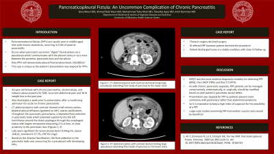
Case Description/Methods: A 43-year-old female with chronic pancreatitis, alcohol abuse, and tobacco abuse presents for SOB, recurrent abdominal pain and 30 lb weight loss in the past year. She was discharged a week prior to presentation after a month-long admission for acute on chronic pancreatitis. A CT abdomen/pelvis with contrast showed small volume ascites, bilateral pleural effusions (greatest on left), coarse calcifications throughout the pancreatic parenchyma, a lobulated fluid collection in pancreatic body which extended superiorly into the left hemithorax around the distal esophagus through the esophageal hiatus with largest component measuring 2.3 x 4.4cm, in close proximity to the pancreatic duct (Image1). Labs were significant for serum procalcitonin 0.04ng/mL, lipase 104U/L, hematocrit 27.7%, CRP 45.1mg/L. Based on the Atlanta Classification, the fluid collection in the pancreatic body was concerning for a pseudocyst with developing PPFs. Thoracic surgery declined surgery. GI offered ERP however patient declined the procedure. She was discharged home in a stable condition with close GI follow-up.
Discussion: In a systematic review, MRCP was the most sensitive diagnostic modality for detecting PPF (80%), then ERCP (78%) and then CT (47%) (1). As with chronic pancreatic sequelae, PPF patients can be managed conservatively, endoscopically, or surgically. Management should be modified based on each patient’s pancreatic ductal defect. Large-scale studies examining PPF intervention success rates would be beneficial. Our patient’s presentation was atypical for PPF as patients present more commonly with pulmonary rather than abdominal symptoms, the opposite of her presentation. So it is imperative to keep a high index of suspicion for the possibility of a PPF.

Disclosures:
Gina Moon, MD1, Ahmad Basil Nasir, MD2, Muhammad Taha Khan, MD3, Tokunbo Ajayi, MD2, Amir Rumman, MD1. P2898 - Pancreaticopleural Fistula: An Uncommon Complication of Chronic Pancreatitis, ACG 2023 Annual Scientific Meeting Abstracts. Vancouver, BC, Canada: American College of Gastroenterology.
1University of Oklahoma Health Sciences Center, Oklahoma City, OK; 2University of Oklahoma College of Medicine, Oklahoma City, OK; 3Oklahoma University Health Sciences Center, Oklahoma City, OK
Introduction: Pancreaticopleural fistulas (PPFs) are usually seen in middle-aged men with chronic alcoholism, occurring in 0.4% of cases of pancreatitis. It occurs when pancreatic secretions “digest” fascial planes via a pseudocyst which communicates with the pleural cavity or via a tract between the posterior pancreatic duct and the pleura. Although there is no defined cutoff value, only PPFs will demonstrate pleural fluid amylase levels >50,000U/L. This case is unique as the patient’s presentation was atypical for PPFs.
Case Description/Methods: A 43-year-old female with chronic pancreatitis, alcohol abuse, and tobacco abuse presents for SOB, recurrent abdominal pain and 30 lb weight loss in the past year. She was discharged a week prior to presentation after a month-long admission for acute on chronic pancreatitis. A CT abdomen/pelvis with contrast showed small volume ascites, bilateral pleural effusions (greatest on left), coarse calcifications throughout the pancreatic parenchyma, a lobulated fluid collection in pancreatic body which extended superiorly into the left hemithorax around the distal esophagus through the esophageal hiatus with largest component measuring 2.3 x 4.4cm, in close proximity to the pancreatic duct (Image1). Labs were significant for serum procalcitonin 0.04ng/mL, lipase 104U/L, hematocrit 27.7%, CRP 45.1mg/L. Based on the Atlanta Classification, the fluid collection in the pancreatic body was concerning for a pseudocyst with developing PPFs. Thoracic surgery declined surgery. GI offered ERP however patient declined the procedure. She was discharged home in a stable condition with close GI follow-up.
Discussion: In a systematic review, MRCP was the most sensitive diagnostic modality for detecting PPF (80%), then ERCP (78%) and then CT (47%) (1). As with chronic pancreatic sequelae, PPF patients can be managed conservatively, endoscopically, or surgically. Management should be modified based on each patient’s pancreatic ductal defect. Large-scale studies examining PPF intervention success rates would be beneficial. Our patient’s presentation was atypical for PPF as patients present more commonly with pulmonary rather than abdominal symptoms, the opposite of her presentation. So it is imperative to keep a high index of suspicion for the possibility of a PPF.

Figure: Image 1: Panels A and B - CT abdomen/pelvis with contrast demonstrating large pseudocyst extending from body of pancreas to the lower chest
Disclosures:
Gina Moon indicated no relevant financial relationships.
Ahmad Basil Nasir indicated no relevant financial relationships.
Muhammad Taha Khan indicated no relevant financial relationships.
Tokunbo Ajayi indicated no relevant financial relationships.
Amir Rumman indicated no relevant financial relationships.
Gina Moon, MD1, Ahmad Basil Nasir, MD2, Muhammad Taha Khan, MD3, Tokunbo Ajayi, MD2, Amir Rumman, MD1. P2898 - Pancreaticopleural Fistula: An Uncommon Complication of Chronic Pancreatitis, ACG 2023 Annual Scientific Meeting Abstracts. Vancouver, BC, Canada: American College of Gastroenterology.
