Tuesday Poster Session
Category: Biliary/Pancreas
P2901 - Bouveret Syndrome: A Rare Cause of Gastric Outlet Obstruction
Tuesday, October 24, 2023
10:30 AM - 4:00 PM PT
Location: Exhibit Hall

Has Audio
- MG
Miles Graves, MD
United Hospital Center
Bridgeport, West Virginia
Presenting Author(s)
Miles Graves, MD1, Kimberly Fairley, DO2
1United Hospital Center, Bridgeport, WV; 2West Virginia University-United Hospital Center, Bridgeport, WV
Introduction: We present a case of Bouveret Syndrome in an 88-year-old male who presented with chief complaints of decreased appetite, nausea, emesis, and abdominal distension of three days in duration. Bouveret Syndrome occurs when a bilioduodenal fistula forms allowing passage of a gallstone into the duodenum that results in gastric outlet obstruction. Bouveret syndrome is extremely rare with only 315 cases reported from 1967 through 2016.
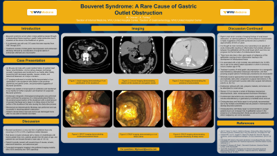
Case Description/Methods: Our patient was an 88-year-old male with a past medical history significant for systolic heart failure with an ejection fraction of 20%, atrial fibrillation, chronic kidney disease, and benign prostatic hyperplasia who presented with decreased appetite, nausea, emesis, and abdominal distension that had been ongoing for 3 days. CT imaging was performed and demonstrated a 4 cm calcification in the duodenum (image 1), dilation of the proximal duodenum and gastric lumen (image 2), and pneumobilia (image 3) suggestive of Bouveret syndrome. He underwent ERCP which confirmed the presence of a cholecystoduodenal fistula in the proximal second portion of the duodenum with a small amount of purulent discharge (image 4) and a large obstructing gallstone in the third portion of his duodenum that was causing his obstructive process (image 5). Mechanical and electrohydraulic lithotripsy was performed and was successful in resolving patient's obstruction (image 6).
Discussion: Bouveret syndrome is more common in females, individuals with a history of cholelithiasis with stones greater than 2cm, and in patients with ages greater than 60 years. Patient's present with several days of nausea, emesis, abdominal distension, and abdominal pain. CT is the preferred imaging modality with a 93% sensitivity and 100% specificity. Rigler's triad which consists of imaging findings of small bowel obstruction, an aberrant gallstone, and the presence of pneumobilia which can all be appreciated on CT imaging is highly suggestive of Bouveret syndrome. Preferred treatment consists of gastric decompression via a nasogastric tube and endoscopic intervention. If the stone is small enough, endoscopic retrieval with nets, graspers, baskets, or snares can be attempted. Stones greater than 2.5 cm typically require mechanical, electrohydraulic, laser, or extracorporeal shockwave lithotripsy. If endoscopic interventions are unsuccessful, enterolithotomy or gastrostomy can be performed to surgically facilitate stone removal.

Disclosures:
Miles Graves, MD1, Kimberly Fairley, DO2. P2901 - Bouveret Syndrome: A Rare Cause of Gastric Outlet Obstruction, ACG 2023 Annual Scientific Meeting Abstracts. Vancouver, BC, Canada: American College of Gastroenterology.
1United Hospital Center, Bridgeport, WV; 2West Virginia University-United Hospital Center, Bridgeport, WV
Introduction: We present a case of Bouveret Syndrome in an 88-year-old male who presented with chief complaints of decreased appetite, nausea, emesis, and abdominal distension of three days in duration. Bouveret Syndrome occurs when a bilioduodenal fistula forms allowing passage of a gallstone into the duodenum that results in gastric outlet obstruction. Bouveret syndrome is extremely rare with only 315 cases reported from 1967 through 2016.
Case Description/Methods: Our patient was an 88-year-old male with a past medical history significant for systolic heart failure with an ejection fraction of 20%, atrial fibrillation, chronic kidney disease, and benign prostatic hyperplasia who presented with decreased appetite, nausea, emesis, and abdominal distension that had been ongoing for 3 days. CT imaging was performed and demonstrated a 4 cm calcification in the duodenum (image 1), dilation of the proximal duodenum and gastric lumen (image 2), and pneumobilia (image 3) suggestive of Bouveret syndrome. He underwent ERCP which confirmed the presence of a cholecystoduodenal fistula in the proximal second portion of the duodenum with a small amount of purulent discharge (image 4) and a large obstructing gallstone in the third portion of his duodenum that was causing his obstructive process (image 5). Mechanical and electrohydraulic lithotripsy was performed and was successful in resolving patient's obstruction (image 6).
Discussion: Bouveret syndrome is more common in females, individuals with a history of cholelithiasis with stones greater than 2cm, and in patients with ages greater than 60 years. Patient's present with several days of nausea, emesis, abdominal distension, and abdominal pain. CT is the preferred imaging modality with a 93% sensitivity and 100% specificity. Rigler's triad which consists of imaging findings of small bowel obstruction, an aberrant gallstone, and the presence of pneumobilia which can all be appreciated on CT imaging is highly suggestive of Bouveret syndrome. Preferred treatment consists of gastric decompression via a nasogastric tube and endoscopic intervention. If the stone is small enough, endoscopic retrieval with nets, graspers, baskets, or snares can be attempted. Stones greater than 2.5 cm typically require mechanical, electrohydraulic, laser, or extracorporeal shockwave lithotripsy. If endoscopic interventions are unsuccessful, enterolithotomy or gastrostomy can be performed to surgically facilitate stone removal.

Figure: Please refer to abstract body text.
Disclosures:
Miles Graves indicated no relevant financial relationships.
Kimberly Fairley indicated no relevant financial relationships.
Miles Graves, MD1, Kimberly Fairley, DO2. P2901 - Bouveret Syndrome: A Rare Cause of Gastric Outlet Obstruction, ACG 2023 Annual Scientific Meeting Abstracts. Vancouver, BC, Canada: American College of Gastroenterology.
